-
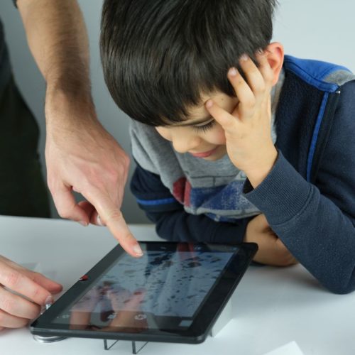
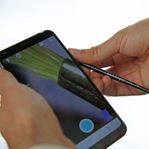
 This is the last generation of the Blips Labkit. With the most powerful lens (Ultra), you can use your phone like a digital microscope: with resolution of 3-4 micron you can see living micro-organisms, or even blood cells on prepared slide. With the extra-thin Macro lens kit (inside the box) you will get powerful lenses to keep always with you, for seeing microscopic details whenever you want.
 -
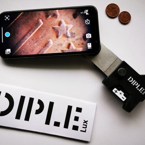
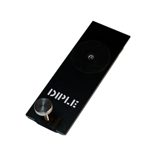
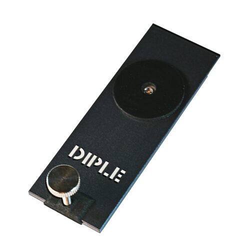
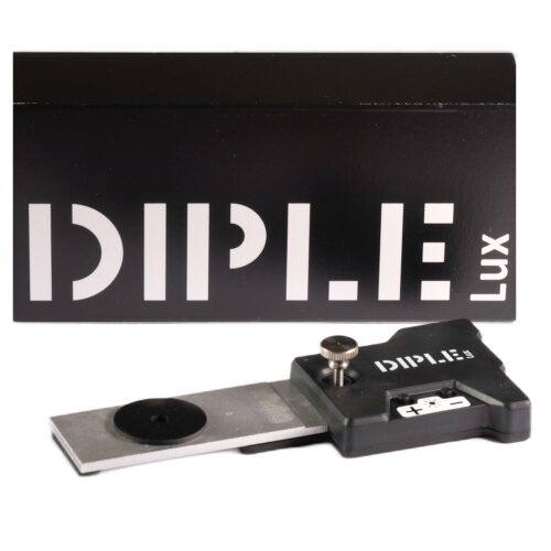 DIPLE LUX is made up of a DIPLE objective LENS, plus a compact EXTRA LIGHT SOURCE for lateral illumination of the samples; a new system specifically designed to work on opaque subjects to observe the tiniest details of their surfaces, out of the DIPLE box. Therefore, DIPLE Lux is the perfect solution to observe opaque subjects. It is, indeed, your new powerful, solid and portable magnifying lens.Download DIPLE Lux instructions hereProduct SKU: D0001
 -
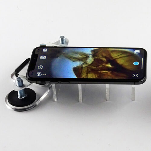
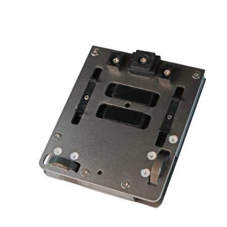 The fine stage: the slide can be shifted by mechanical screws, for a precise search of microscopic targets.
 -
 1 DIPLE Black lens 150x - Optical resolution of 0.7-0.8 micron. With digital zoom, it allows to reach a magnification of about 1000x on the screen of the used device, without pixelation.
 -
- Extremely portable: you can keep it in your wallet
- Clear, high quality shots saved directly on your device
- Very powerful: max resolution of 0.0035 mm (Blips Ultra), 0.005 mm (Blips Micro), 0.01 mm (Blips Macro) and 0.015 mm (Blips Macro Plus)
- Reusable for hundreds of times
- Spare, multi-usable adhesive tape included
- Free App included
-
 1 Diple Grey lens 75x - Resolution of about 1 micron. It allows to reach a magnification of about 500x on the screen of common devices just adding digital zoom, without significant pixelation.
 -
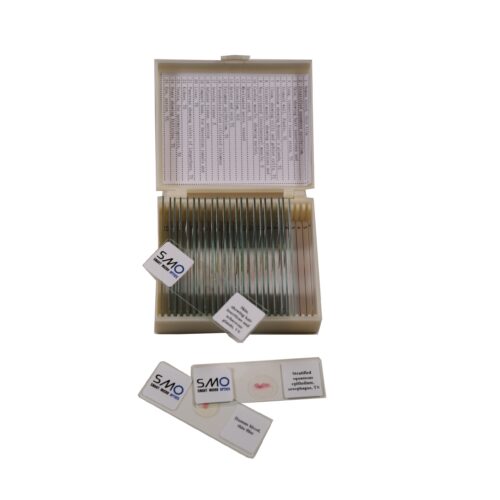 A set of 25 prepared slides for microscopy, with various histology specimens. With a solid plastic carrying box.
- Human blood, thin film
- Stratified squamous epithelium, oesophagus, TS
- Skin, showing hair insertions and sebaceous glands, VS
- Lung, connective tissue stain, TS
- Stomach wall, fundic, VS
- Duodenum showing Brunner's glands, TS
- Ileum, showing villi and goblet cells, TS
- Striated muscle, showing striations and nuclei, LS
- Non-striated (involuntary) muscle, E
- Cardiac muscle, showing nuclei, striations and intercalated discs, LS
- Spinal cord, TS
- Myelinated nerve, teased
- Cerebellum, VLS
- Artery and vein TS
- Columnar epithelium, TS
- Pseudostratified cilliated columnar epithelium, TS
- Olar tissue E
- Adipose tissue, section
- Compact bone, for Haversian canals and lamellae, TS
- Liver, TS
- Pancreas showing islets of Langerhans, TS
- Kidney, TS
- Testis, for spermatogenesis, TS
- Ovary showing follicles, TS
- Eye, entire, VS
-
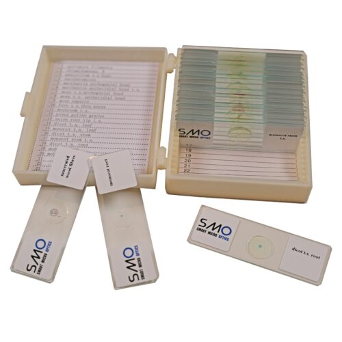 A set of 20 prepared slides for microscopy, with various botany specimens. With a solid plastic carrying box. 01. Spirogyra filaments 02. Chlamydomonas, E 03. Penicillum v.s host 04. Saccharomyces 05. Marchantia archegonial head 06. Marchantia antheridial head v.s. 07. moss v.s.archegonial head 08. moss v.s. antheridial head 09. moss capsule 10. fern v.s.thru.sorus 11. mushroom v.s. 12. pinus pollen grains 13. onion root tip l.s. 14. dicot t.s. leaf 15. monocot t.s. leaf 16. dicot t.s. stem 17. monocot stem t.s. 18. dicot t.s. root 19. monocot root 20. macerated wood fibres


Via Reale 203, 48123 Ravenna, Italia
info@smartmicrooptics.com
SmartMicroOptics srl
P.IVA IT02382790992
© Copyright 2015 –
All Rights Reserved
COMPANY
LEGAL INFO
CUSTOMER SERVICE
Design by Professionalsite